In multiple sclerosis (MS), diffuse degenerative processes in the deep grey matter have been associated with clinical disabilities. We performed a systematic study in MS deep grey matter with a focus on the incidence and topographical distribution of lesions in relation to white matter and cortex in a total sample of 75 MS autopsy patients and 12 controls. In addition, detailed analyses of inflammation, acute axonal injury, iron deposition and oxidative stress were performed. MS deep grey matter was affected by two different processes: the formation of focal demyelinating lesions and diffuse neurodegeneration. Deep grey matter demyelination was most prominent in the caudate nucleus and hypothalamus and could already be seen in early MS stages. Lesions developed on the background of inflammation. Deep grey matter inflammation was intermediate between low inflammatory cortical lesions and active white matter lesions. Demyelination and neurodegeneration were associated with oxidative injury. Iron was stored primarily within oligodendrocytes and myelin fibres and released upon demyelination. In addition to focal demyelinated plaques, the MS deep grey matter also showed diffuse and global neurodegeneration. This was reflected by a global reduction of neuronal density, the presence of acutely injured axons, and the accumulation of oxidised phospholipids and DNA in neurons, oligodendrocytes and axons. Neurodegeneration was associated with T cell infiltration, expression of inducible nitric oxide synthase in microglia and profound accumulation of iron. Thus, both focal lesions as well as diffuse neurodegeneration in the deep grey matter appeared to contribute to the neurological disabilities of MS patients.
Grey matter (or gray matter) is a major component of the central nervous system, consisting of neuronal cell bodies, neuropil (dendrites and myelinated as well as unmyelinated axons), glial cells (astroglia and oligodendrocytes) and capillaries. Grey matter is distinguished from white matter, in that grey matter contains numerous cell bodies and relatively few myelinated axons, while white matter is composed chiefly of long-range myelinated axon tracts and contains relatively very few cell bodies. The color difference arises mainly from the whiteness of myelin. In living tissue, grey matter actually has a very light grey color with yellowish or pinkish hues, which come from capillary blood vessels and neuronal cell bodies.
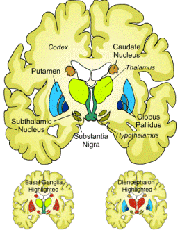
Grey matter is distributed at the surface of the cerebral hemispheres (cerebral cortex) and of the cerebellum (cerebellar cortex), as well as in the depths of the cerebrum (thalamus; hypothalamus; subthalamus, basal ganglia - putamen, globus pallidus, nucleus accumbens; septal nuclei), cerebellar (deep cerebellar nuclei -dentate nucleus, globose nucleus, emboliform nucleus, fastigial nucleus), brainstem(substantia nigra, red nucleus, olivary nuclei, cranial nerve nuclei) and spinal grey matter (anterior horn, lateral horn, posterior horn).
Lesions in the grey matter can result from direct attack of the grey matter or could results as a consequence of events in the white matter. In this study they looked a grey matter structures away from the surface of the brain (=cortex), which had more inflammation than the cortex but less than the white matter.
Is that there was inflammation in the vicinity of damage. This was associated with frank destructive inflammation and also defuse damage. It has been reported previously (click here) that this may be secondary to damage to white matter tracts that project to the grey matter.
Histochemical and MRI studies have demonstrated that MSers have abnormal deposition of iron in both gray and white matter structures. Iron could partake in pathogenesis by various mechanisms, e.g., promoting the production of reactive oxygen species and enhancing the production of proinflammatory cytokines, etc. etc., etc.

Oxidative injury (such as free radicals, hydorgen peroxide, superoxide= Oxygen with three oxygen molecules..into normally has two in oxygen gas, nitric oxide) can be amplified by iron, which accumulates with age in the normal human brain and is liberated from oligodendrocytes and myelin during active demyelination.

Pattern of cortical, white matter and deep grey matter demyelination in Acute MS (ACMS), Relapsing MS (RRMS) and secondary (SPMS)and PPMS. The extent of evaluated demyelination was marked in green for white matter demyelination, in yellow for white matter remyelination, in red for cortical demyelination and in blue for DGM demyelination.
Look at the depth of the sulci (brain folding) in SPMS and PPMS which are enlarged as a consequence of nerve loss.
The deep grey matter structures are profoundly involved in the disease progression of MS patients and appears to contribute significantly to the global accumulation of disability in MS patients. In addition to focal demyelination, a diffuse neurodegenerative process occurs in the DGM and is associated with clinical disability.